
Visual Representations and Perceptions
of Lipoproteins and Atherosclerosis
- Elisa Campos
_______________________________
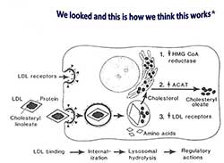
Fig. 13. Sequential steps in the LDL receptor
pathway of mammalian cells, 1979 

Fig. 14. The LDL receptor pathway
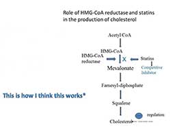
Fig. 15. The Role of HMG-CoA reductase
and statins in the synthesis of cholesterol
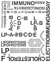
Fig. 16. Al. Alaupovic, A tribute to
lipoprotein particles, 1990
The Search for the Fundamental Deficiency in Familial Hypercholesterolemia
Two researchers, Michael Brown (b.1941) and Joseph Goldstein (b.1940), first met during their internship at Massachusetts General Hospital in Boston (19661968) and share a great interest in the molecular basis of the disease, i.e. in the process of molecularization. Both later went on to the NIH. Brown and Goldstein used to discuss patients with abnormalities of lipid metabolism, in particular patients’ homozygous for familial hypercholesterolemia. It was at the NIH that Goldstein, originally from Dallas, asked Brown to go to the University of Texas Health Science Center in Dallas to work on the genetic regulation of cholesterol metabolism. They hoped to clarify the biochemical mechanism by which a single gene increases cholesterol levels in this rare patient group of hypercholesterolaemic homozygotes on a molecular level (fig. 13).
The team had an experimental system which they believed to be a useful approach in the search for the fundamental deficiency, apparently genetically defined in FH, and causing a defect in the regulation of cholesterol synthesis at the level of HMG-CoA reductase. This suggested the hypothesis of a receptor on the membrane surface binding to an LDL receptor. As a result of this binding, a second messenger would be generated suppressing the activity of the reductase.
Experimentation reflects the scientist’s desire to observe or experience the operation of cause and effect in order to verify or reject a hypothesis. Brown and Goldstein used the technique of growing cells in culture, which was relatively new in 1972. The notion that cultured cells might allow pinpointing of metabolic errors was still newer. Thus they introduced cell biology into the study of lipoproteins, again as a means of making phenomena illustrate themselves. An illustration was worked up to make its visualization and interpretation as direct as possible (fig. 14).
Akira Endo and the Discovery of Statins
Knowledgeable about cholesterol synthesis and also aware that the limiting step of the synthesis is the one in which HMG-CoA reductase intervenes, Akira Endo (b.1933) thought that an inhibitor of this enzyme would be an effective agent in reducing cholesterol levels. And because the company where he was in 1971 was carrying out studies on fungi, he speculated that just as some species inhibit bacterial growth, others could inhibit the synthesis of cholesterol.
Over a two-year period, Endo’s team tested more than 6,000 fungi for their ability to block cholesterol synthesis. In 1973 they finally found one strain of fungus that produced an active compound. The fungus belonged to the same genus that produced penicillin, the reason some people call the statins penicillin for the heart. Figure 15 shows how Brown and Goldstein’s work strongly supported Endo’s studies and his idea of developing HMG-CoA reductase inhibitors.
It is this use of Nature that sets their work apart, and when communication of an understanding of Nature through images allows the scientist to say, This is how it works, the picture, to a certain extent, becomes the theory; subsequently, research and experimental practice will be stimulated. According to Chadarevian and Kamminga [6], it is rare to have a convergence of etiology, diagnosis and therapy; i.e., knowing the mechanisms of disease and the diagnostic at a molecular level does not imply that a molecular cure is available. In the case of hypercholesterolemia and of statins, the mechanism of the action of the therapeutic molecule is not the only known factor, and statins have few side effects, being considered safer than aspirin [7].
Scientists’ images always aim to give what they consider to be the most rational version of phenomena. However, how specific traits should be visualized and how to pick the perfect image depend to a large degree upon pragmatic categories and local factors within individual laboratories and research groups, as well as on editorial decisions and promotional value. Experimental scientists work with a combination of action, instruments, objects and procedures as well as words, that is, with a significant non-verbal component of communication.
Visualization is thus an integral part of the understanding and evolution of new scientific concepts and boundaries. The images made for pragmatic and heuristic purposes in the laboratory evolve into those that are chosen for posters and lecture presentations, images accompanying article submissions, and finally those that will be selected for public presentation and communication. We conclude, simply, with a work of art by Alexandra Alaupovic entitled A Tribute to Lipoprotein Particles (fig. 16).
[6] Chadarevian, S., Kamminga, H., Introduction, Molecularizing Biology and Medicine: New Practices and Alliances, 1910s-1970s, eds. Chadarevian, S. and Kamminga, H., Amsterdam, Harwood Academic Publishers, 1998, pp. 1-16.
[7] Steinberg, D., The Cholesterol Wars: The Skeptics vs. The Preponderance of Evidence, Amsterdam, Elsevier, 2007, p. 199.


